
Prev
Next
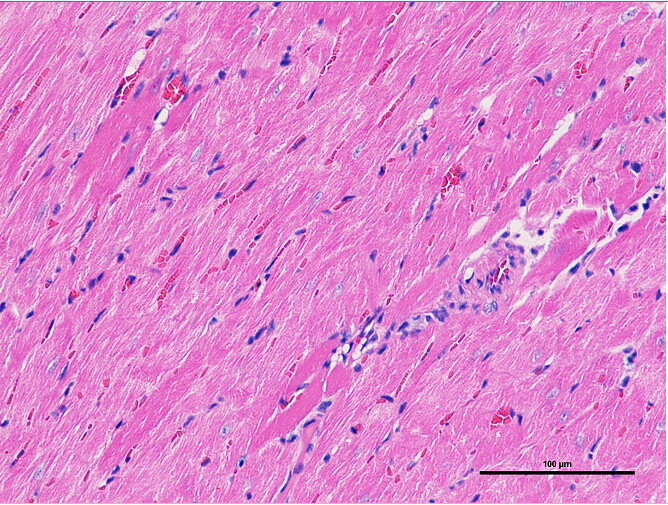
detailed informationHE staining, which is the hematoxylin-eosin staining method, is the most commonly used morphological staining method. On HE stained sections of better quality, the general morphological characteristics of various tissues or cells can be observed. Hematoxylin staining solution is alkaline, which mainly makes the chromatin in the nucleus and ribosomes in the cytoplasm to be purple blue; eosin is an acid dye, which mainly makes the components in the cytoplasm and extracellular matrix red.
 [Operating procedures] 1. Bake paraffin sections in an oven at 60 ° C for 1 to 2 hours; 2. Paraffin section conventional xylene, ethanol dewaxing to water, 3. Hematoxylin staining for 10 minutes, 4. Rinse under running water to remove residual color, 5.0.7% ethanol hydrochloride differentiation for several seconds, 6. Rinse under running water, the slices turn blue for about 15 minutes, 7.95% ethanol for 30 seconds, 8. Alcoholic eosin dyeing for 30 seconds, 9.I 95% ethanol for 30 seconds, 10.II 95% ethanol for 30 seconds, 11.I 100% ethanol for 30 seconds, 12.II 100% ethanol for 30 seconds, 13. Xylene phthalate carbonate for 30 seconds, (1: 4 1-xylene carbonate 4) 14.I xylene for 30 seconds, 15.II xylene for 30 seconds, 16. Neutral gum seals. [Experimental results]: The nucleus is blue under microscope; the cytoplasm is generally red; collagen fibers are red to varying degrees, red blood cells are bright red; mucin is light blue; calcified tissue is dark blue. [Problems and solutions in the experiment]: 1. Nuclei are pale and dim Reason: There may be 3 factors affecting this type of problem. The residence time of the sections in the hematoxylin staining solution is too short; the hematoxylin staining solution is over-oxidized, loses its ability to stain, and can no longer be used; the differentiation step takes too long. Countermeasure: restain the section. Properly extend the hematoxylin staining time; replace the hematoxylin staining solution; shorten the differentiation time. Staining and differentiation time can be explored through microscopic observation. 2. The nuclei are overstained, that is, the nuclei are too dark blue, and they are connected to the surrounding nuclei, and the outline is not clear. Reason: There may be 3 influencing factors for this type of problem, that is, the section stays too long in the hematoxylin staining solution, the section is too thick, and the differentiation step is too short. Countermeasures: Decolorize, bleach, and restain the sections, and make appropriate adjustments to the staining and differentiation time in combination with microscopic observation. If it is determined that the nuclei are overstained due to the section being too thick, resection is required. Eosin Reason: Eosin solution may be old and lose its ability to stain; or it may be dehydrated in ethanol for too long after staining with eosin. Countermeasures: Decolorize, bleach, and restain the sections, and make appropriate adjustments to the staining and differentiation time in combination with microscopic observation. If it is determined that the nuclei are overstained due to the section being too thick, resection is required. |
Contact Us
|